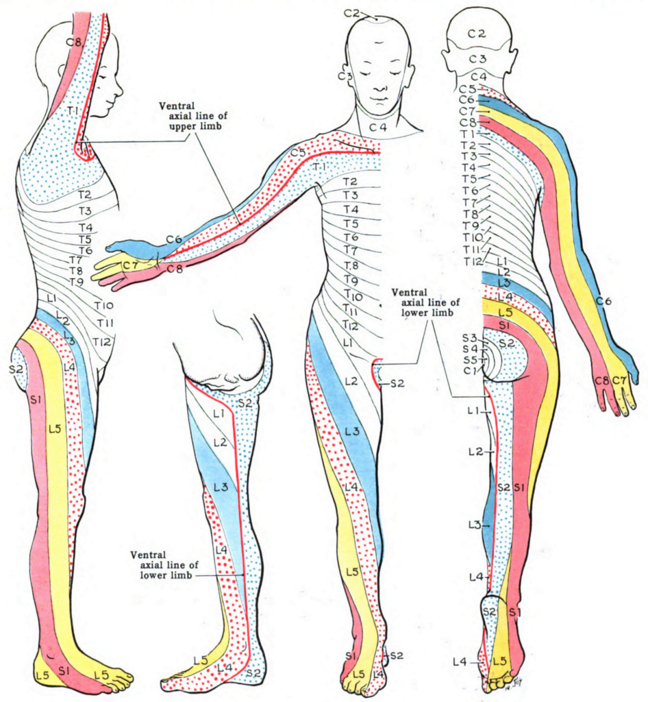
Dermatomes Chart Leg – The term “dermatome” is a mix of two Ancient Greek words; “derma” meaning “skin”, and “tome”, indicating “cutting” or “thin section”. It is a location of skin which is innervated by the posterior (dorsal) root of a single spine nerve. As posterior roots are organized in sectors, dermatomes are. This is why the term “dermatome” refers to the segmental innervation of the skin.
Dermatome Anatomy Wikipedia – Dermatome anatomy Wikipedia
Neighboring dermatomes often, if not constantly overlap to some degree with each other, as the sensory peripheral branches corresponding to one posterior root generally surpass the limit of their dermatome. As such, the thin lines seen in the dermatome maps are more of a medical guide than a real boundary. Dermatomes Chart Leg
This suggests that if a single spinal nerve is affected, there is most likely still some degree of innervation to that segment of skin originating from above and listed below. For a dermatome to be completely numb, typically 2 or 3 surrounding posterior roots need to be affected. In addition, it’s crucial to note that dermatomes undergo a big degree of interindividual variation. A visual representation of all the dermatomes on a body surface chart is described as a dermatome map. Dermatomes Chart Leg
Dermatome maps
Dermatome maps depict the sensory circulation of each dermatome across the body. Clinicians can assess cutaneous experience with a dermatome map as a method to localize lesions within main worried tissue, injury to particular back nerves, and to identify the degree of the injury. Several dermatome maps have actually been developed throughout the years but are often contrasting.
The most frequently utilized dermatome maps in major textbooks are the Keegan and Garrett map (1948) which leans towards a developmental interpretation of this concept, and the Foerster map (1933) which associates better with medical practice. This post will examine the dermatomes utilizing both maps, determining and comparing the major differences between them.
Why Are Dermatomes Important?
To comprehend dermatomes, it is important to comprehend the anatomy of the spinal column. The spine is divided into 31 sections, each with a pair (right and left) of anterior and posterior nerve roots. The kinds of nerves in the posterior and anterior roots are different.
Anterior nerve roots are responsible for motor signals to the body, and posterior nerve roots get sensory signals like discomfort or other sensory signs. The posterior and anterior nerve roots combine on each side to form the back nerves as they leave the vertebral canal (the bones of the spine, or foundation).
